Generating X-rays
July 5, 2011
Gen-X would have been a good name for an
X-ray equipment company. Unfortunately, the term has been usurped as the name of the generation born after the
baby boom of which I was a part. Gen-X a fuzzy term, meaning different things to different people. It's most commonly applied to people born in the 1960s and 1970s. They missed
Sputnik, but many of them experienced the
first manned moon landing. That's quite a leap in technology in just one generation!
Like most
materials scientists, I did a lot of work with X-rays. There was routine
powder diffraction analysis of
phases, but there was also the more exotic
linewidth determination by a double-crystal
diffractometer to determine the quality of
single crystals. Having worked with X-rays for many years, I have two interesting
anecdotes.
The first comes from the time when my fellow
graduate students and I were first learning X-ray technique. Since we were working with X-rays, we each needed X-ray
dosimeter badge. One of the students didn't quite understand the badge concept, so he asked how a small piece of plastic could protect him from the X-rays.
Then there was the month when all the dosimeter badges in our laboratory showed an exposure. We checked all three machines in the room, and there were no stray X-rays emitted by any. As it turned out, someone who was working with a harmless
tracer chemical had left a vial of it on the shelf where everyone kept their exposure badges.
The generation of X-rays is quite simple. So simple, in fact, that equipment not designed to emit X-rays can emit them.
Electrons of sufficiently high energy impacting materials will generate X-rays. All you need is a source of high energy electrons and a target. There aren't that many
cathode ray tubes out there, now, but the introduction of
color television caused a problem.
Color television tubes incorporate a
shadow mask to define the color fields on the
phosphor screen, and electrons impacting on this screen generate X-rays. The electrons impacting on the phosphor are less of a problem, since the phosphor
elements needed higher electron energies to produce X-rays. Not surprisingly, such emissions are
limited by law, but CRT televisions are now a rarity.
Wilhelm Röntgen was awarded the first
Nobel Prize in Physics in 1901 for his discovery of X-rays in 1895. X-rays soon became a common tool for
scientific and
medical investigations. By 1933, a patent was issued for the first
rotating anode X-ray tube for generating extremely high X-ray power.[1] One of my aunts had X-ray treatments for
acne in the 1930s, a medical procedure that's not currently recommended!

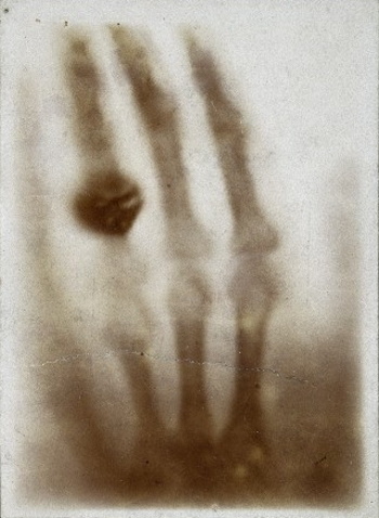
The first medical X-ray.
Hand mit Ringen: Wilhelm Röntgen's x-ray of his wife's hand, taken on December 22, 1895.
(Via Wikimedia Commons)
Stefan Kneip of the
Blackett Laboratory,
Imperial College (London, UK) has written a review of novel methods of X-ray generation in a recent issue of
Nature.[2] One method of special interest is that pioneered at
Seth Putterman's laboratory at UCLA.
Putterman is a wellspring of novel ideas,[3-4] and I've mentioned his work in a
previous article (Pyroelectric Energy Harvesting, October 15, 2010). Putterman's group discovered that the
electrostatic charge developed when something as common as unrolling
pressure-sensitive tape will generate X-rays.[5-8] This work made the cover of the October 23, 2008, issue of Nature.[5]
The principle is called
triboluminescence, and Putterman's
UCLA team found that peeling common adhesive tape in a moderate
vacuum will produce emission throughout the
electromagnetic spectrum, from
radio through X-rays. The X-rays were concentrated in 100-
mW pulses that were correlated with the stick–slip action involved in the peeling. The X-ray emission was found to occur at a gap between the separating faces of the tape of 30 - 300
μm with an emission peak at 15-
keV. They were able to use the emission for X-ray imaging.[3] In homage to Röntgen, the Nature cover image was of a human finger imaged using X-rays from
Scotch-brand tape.
The study was done with a device that unspooled tape at 1.3 inches per second.[6] One observation was that
duct tape did not produce X-rays.[7] The production of X-rays required the tape to be unspooled in a vacuum, since the
mean free path of electrons is too short in air, or
humidity short-circuits the
electric field.[7]
In a follow-up study published in May, 2011,[9] the UCLA group improved their apparatus to have
silicone and a
metal-filled
epoxy repeatedly contact each other in vacuum. The apparatus uses a
solenoid to make, and break, contact twenty times a second, but they are investigating a
piezoelectric actuator that will operate at 300
Hz.[2]
Other material couples may be better at creating triboelectricity.[2]
According to calculations, pulling apart this silicone-epoxy couple generates 10
10 electrons per cm
2.[2] The machine was able to produce X-rays from
molybdenum and
silver target materials at a rate of 10
5 photons per contact cycle. The electron energy was 40 keV. They predict that an X-ray flux of up to 10
8 photons per second would be possible, and such a device could be an inexpensive source of X-rays.
References:
- Albert Bouwers, "X-Ray Tube," US Patent No. 1,933,005, Oct 31, 1933
- Stefan Kneip, "Applied physics: A stroke of X-ray," Nature, vol. 473, no. 7348 (May 26, 2011), pp. 455-456.
- Seth Putterman, James K. Gimzewski, Brian B. Naranjo, "High energy crystal generators and their applications," US Patent No. 7,741,615, June 22, 2010.
- Brian Naranjo, James Gimzewski, Seth Putterman, "Method And Apparatus For Generating Nuclear Fusion Using Crystalline Materials,"US Patent Application No. 11/745,556, Publication number: US 2008/0142717 A1, May 8, 2007.
- Carlos G. Camara, Juan V. Escobar, Jonathan R. Hird and Seth J. Putterman, "Correlation between nanosecond X-ray flashes and stick–slip friction in peeling tape," Nature, vol. 455, no. 7216 (October 23, 2008), pp. 1007-1148.
- Thomas H. Maugh II, "UCLA researchers' surprising finding could lead to applications in medicine, other fields," LA Times, October 25, 2008.
- Kenneth Chang, "Scotch Tape Unleashes X-Ray Power," The New York Times, October 28, 2008
- Dave Bullock, "Gallery: Take an X-Ray With Your Office Sticky Tape," Wired, October 22, 2008 .
- J. R. Hird, C. G. Camara, and S. J. Putterman, "A triboelectric x-ray source," Applied Physics Letters, vol. 98, no. 13 (March 28, 2011), Document No. 133501 (3 pages).
Permanent Link to this article
Linked Keywords: Gen-X; X-ray; baby boom; Sputnik; Apollo 11; first manned moon landing; materials science; materials scientist; powder diffraction; crystal phase; X-ray crystallography; linewidth; diffractometer; single crystal; anecdote; graduate student; dosimeter badge; tracer chemical; electron; cathode ray tube; color television; shadow mask; phosphor; chemical element; Wilhelm Röntgen; Nobel Prize in Physics; scientific; medical; rotating anode X-ray tube; acne; Wikimedia Commons; Stefan Kneip; Blackett Laboratory; Imperial College (London, UK); Nature; Seth Putterman; electrostatic charge; pressure-sensitive tape; triboluminescence; University of California, Los Angeles; UCLA; vacuum; electromagnetic spectrum; radio; milliwatt; mW; micometer; μm; electronvolt; keV; Scotch-brand tape; duct tape; mean free path; humidity; electric field; silicone; metal; epoxy; solenoid; piezoelectric actuator; hertz; Hz; triboelectric series; molybdenum; silver; photons; US Patent No. 1,933,005, Oct 31, 1933; US Patent No. 7,741,615; US Patent Application No. 11/745,556, Publication number: US 2008/0142717 A1.